Full Length Research Paper - (2023) Volume 18, Issue 1
Immunity Indices Improve In Children With Cerebral Palsy Following Laser Acupuncture Biostimulation
Rowida S El Said*, Mona Morsy, Faten H Abdelazeim and Ola A Dabbous*Correspondence: Rowida S El Said, Department of Physical therapy, Benha Fever Hospital, Benha, Egypt, Email:
2Medical Department, National Institute of Laser Enhanced Sciences (NILES), Cairo University, Giza, Egypt
Department of Physical Therapy for Pediatrics, Faculty of Physical Therapy, Cairo University and Dean, Faculty of Physical Therapy, October 6 Universi, Egypt
3Medical Department, National Institute of Laser Enhanced Sciences (NILES), Cairo University, Giza, Egypt
Received: 05-Jan-2023 Accepted: 06-Feb-2023 Published: 13-Feb-2023
Abstract
One of the most common diseases that is the reason of severe neurological disability is Cerebral Palsy (CP). CP worldwide prevalence is average 1-4 per 1000 live births. The purpose of study was to confirm the impact of laser acupuncture therapy in improvement of immunity indices in children with spastic CP by rising NK cells count. From the pediatric outpatient clinic of the National Institute of Laser Enhanced Sciences (NILES) and the physical therapy outpatient clinic at Benha Fever Hospital, 40 children with spastic CP aged 1 to 3 years of both genders were recruited. They were split into two groups of equal number at random: The physical therapy sessions for Group A (the control group) consisted of one hour each session, three times each week, for a total of 12 weeks. In addition to their regular physical treatment, participants in Group B (the study group) also underwent 780 nm Diode laser acupuncture with an output power of 50 mw three times weekly for a total of 12 sessions. All children in both groups had their NK cells (CD16 CD56) measured by flow cytometry using “BD Facscalibur” with two different colors before and after treatment. Statistical tests were performed on the data. Counts of NK cells (CD16, CD56) differed significantly between group A and group B after treatment, with group B having a higher total. The results of this study showed that the number of NK cells in the children with spastic CP increased after they had laser acupuncture treatment.
Keywords
Spastic cerebral palsy. Low level laser therapy 780nm. Laser Acupuncture. Immunity. Natural killer cells. Flow cytometry.
Introduction
The foremost of motor disability in children is caused by CP. Recently, it has been sorted out a combination of lasting abnormalities of development and position that lead to confined activity which are attributed to static disturbing impacts that occurred during prenatal or newborn brain development [1]. Often, CP occurs during pregnancy, during labor or shortly after birth as reason of mal development or harmful of the brain parts that considered the leadership of movement, balance, and posture [2]. In CP children; there is impairment of regulation of immune that caused via auto-reaction against damaged nerve tissue [3]. Because of the tight connection between the neurological system and the immune system, CP children are vulnerable to physiological regulation of restrictions of blood flow of the nervous tissue [4].
NK cells are famous for their cytotoxic feature against virus infected or changed cells, besides their feature of cytokine generation capabilities [5]. Blood pictures taken before and after low-level laser therapy (LLLT) treatment showed a considerable increase in CD45 lymphocytes and NK cells (CD16 CD56) [6].
Laser acupuncture therapy (LA) is the utilized of non-thermal, low-intensity laser illumination to fortify needle therapy points-has gotten to be more common among needle therapy professionals in later a long time. LA is advanced as a more secure pain-free elective to conventional needle therapy, with negligible unfavorable impacts and more noteworthy flexibility [7].
Acupuncture increases NK cells counts by bolstering the neurotransmitter network and the immune system's transmitter [8]. It is defined by the world health organization 361 acupoints [9]. Stomach 36 (ST36) in Chinese zusanli is the most commonly needle in acupuncture clinics which used to enhance the function of Immunity system [10]. Although, there were previous findings discussed several modalities to improve immunity, there were fewer studies that used laser therapy for immunity improvements. In the current study, we looked at how laser acupuncture affected immune measures for children who have spastic CP.
Subjects
A total of "forty children with spastic CP" of both sexes were enrolled in the clinical study, which took place between January 2021 and December 2021 at the pediatric outpatient clinic of the National Institute of Laser Enhanced Sciences (NILES) and the physical therapy outpatient clinic of Benha Fever Hospital
Criteria for all participants
These were the criteria's selected children: First, there was a referral from a pediatrician confirming a diagnosis of spastic CP. Second, they were between the ages of one and three. Third, the gross motor function classification scheme placed children with spastic CP at level 4 (GMFCS). Finally, using the Modified Ashworth Scale, their muscle tone was anywhere from a 1 to a 1+.
For the following reasons, children were not included in the study:
• If you have any kind of knee deformity
• Having epilepsy.
• Having a problem with your back or having a deformity there.
• Medically unstable, having a history of instability and comorbid conditions such as cardiovascular disease and respiratory diseases like pneumonia.
• Being treated with antibiotics or steroids.
• A genetic disorder.
• Being diagnosed with cancer and undergoing irradiation treatment.
• Following an explanation of the study to the parents of participating children, the National Institute of Laser Enhanced Sciences and Benha Fever Hospital’s respective scientific and ethical committees required the parents to sign an informed permission form before the research could begin.
Design of the study
Subjects were randomly randomized into two groups of similar count and duration of treatment for 12 weeks
• Those in Group A (the controls) attended physical therapy only for a total of 3 (sessions) x 1(hr.) / week.
• To supplement their regular physical therapy, participants in Group B (the study group) also underwent laser acupuncture at immunity acupoint stomach 36 (ST36 in Chinese Zusanli) and additional acupuncture points BL18 (Gan Shu), BL20 (Pi Shu), and BL23 (Shen Shu) three times weekly.
Procedures
This study’s methodology was separated into two major sections:
Evaluation Techniques
For this investigation, natural killer cell expression (CD16 CD56) was measured in all children before and after the study using flow cytometry (“BD Facscalibur”) with two different colours. The levels of natural killer cells in each children’s blood were assessed using an examination of CD16CD56 expression (arterial blood sample was by the drawn from radial artery by the chemist).
Treatment techniques
Physical therapy application
Both a control and a study group participated using a Neurodevelopmental Treatment (NDT) based approach, the program included stretching, range of motion, strengthening, flexibility, and endurance exercises with the goal of enhancing motor functioning and coordination. The program’s primary focus was on enhancing participants’ ability to stand and walk more steadily and confidently.
Acupuncture with a laser
All children in Group B were given laser acupuncture treatments using a Diode Laser device (Laser Level System) to boost their immune system’s acupuncture sites. Table 1 displays the utilized laser's parameters (Table 1).
| Irradiation parameter | Unite of measurement |
|---|---|
| Type of Laser Acupuncture | Low Level Laser Acupuncture |
| Wavelength | 780nm. |
| Mode | continuous wave |
| Beam spot diameter | 1cm. |
| Laser's frequency | 3.8 ×10¹⁴ Hz |
| Output Power | 50 mw. |
| Dose | 3 J /cm2 |
| Treatment time /point | 60 sec. |
| Number of point | 8 points |
| Treatment interval | 3sessions / week |
• Beam spot diameter of laser device was 4mm which was magnified by lens to reach 1cm.
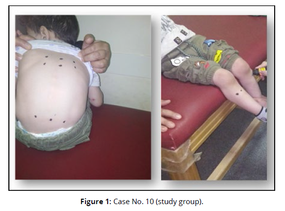
• The probe was placed on the acupoints of the immune system, as depicted in figure 1 (Figure 1).
• Below the patella, on the lateral side of the anterior crest of the bilateral tibial muscles, is the acupoint known as ST36.
• Back lateral to the lower border of the spinous process of the 9th thoracic vertebra on both sides is where BL 18 (Gan Shu) was located.
• Back lateral to the lower border of the spinous process of the 11th thoracic vertebrae on both sides is where BL 20 (Pi Shu) was located.
• A bilaterally located BL 23 (Shen Shu) was identified in the back, on the lateral side of the lower border of the spinous process of the second lumber vertebrae.
Statistical Analysis
The calculated data statistically analyzed and treated to show the mean, standard deviation, and the percentage (for qualitative variables) for all subjects. Paired-t-test used to determine the difference before and after the treatment.
Results
The effects of diode laser acupuncture on indices of immunity were studied in children with spastic cerebral palsy. Data on CD16, CD56, and CD16 CD56 for each child in the two groups were analyzed to get these conclusions (A and B).
CD16
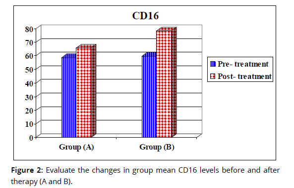
A. Evaluate the changes in group mean CD16 levels before and after therapy (A and B)
Group A’s mean SD pre-treatment value was 58.73 ±14.71, and group A’s post-treatment value was 65.87 ±10.75; there was a statistically significant difference between these two values (p= 0.001), and the percentage change (% change = 12.16) (Table 2). Table 2 shows that between pre- and post-treatment, the mean value of group (B) was 59.85±7.33 (SD), and after treatment, it had increased to 78.4± 7.73 (p0.0001), representing a 31% increase (Figure 2).
Items |
CD16 | |||
|---|---|---|---|---|
| Group (A) | Group (B) | |||
| Pre- Treatment |
Post- Treatment |
Pre- Treatment |
Post- treatment |
|
| x̄± SD | 58.73 ± 14.71 |
65.87 ± 10.75 |
59.85 ± 7.33 |
78.4 ± 7.73 |
| MD | 7.14 | 18.55 | ||
| % of change | 12.16 % | 31 % | ||
| t-value | 3.87 | 18.57 | ||
| p-value | 0.001 | 0.0001 | ||
| Level of Significant | S | S | ||
Evaluate the changes in group mean CD16 levels after therapy (A and B)
After treatment, the mean (SD) values for Group B were 19.02% higher than Group A’s (65.87 ±10.75 vs. 78.4 ±7.73; table 3), yielding a significant difference (p 0.0001) in favor of Group B. (Figure 2 & Table 3).
Items |
CD16 (Post- treatment) |
|
|---|---|---|
| Group (A) | Group (B) | |
| x̄± SD | 65.87 ± 10.75 |
78.4 ± 7.73 |
| MD | 12.53 | |
| % of change | 19.02 % | |
| t-value | 4.23 | |
| p-value | 0.0001 | |
| Level of Significant | S | |
CD56
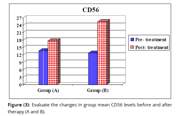
Evaluate the changes in group mean CD56 levels before and after therapy (A and B)
Group (A) pre-treatment's mean SD value was 13.66± 4.8, and the posttreatment mean SD value was 17.68± 5.39; there was a statistically significant difference between these two values (p= 0.0001), and the percentage change was 29.42%. Mean values before and after treatment for group (B) were 12.8± 3.49 and 25.33 ±4.26, respectively (Table 4), with a statistically significant difference between the two (p= 0.0001), and a correspondingly large percentage of change (97.26%) (Figure 3).
Items |
CD56 | |||
|---|---|---|---|---|
| Group (A) | Group (B) | |||
| Pre- Treatment |
Post- Treatment |
Pre- treatment |
Post- treatment |
|
| x̄± SD | 13.66 ± 4.8 |
17.68 ± 5.39 |
12.8 ± 3.49 |
25.33 ± 4.26 |
| MD | 4.02 | 12.45 | ||
| % of change | 29.42 % | 97.26 % | ||
| t-value | 6.07 | 21 | ||
| p-value | 0.0001 | 0.0001 | ||
| Level of Significant | S | S | ||
Evaluate the changes in group mean CD56 levels after therapy (A and B)
Group B showed a statistically significant difference (p= 0.0001) in posttreatment mean SD values (17.68± 5.39 vs. 25.33± 4.26; table 5), and their percentage of change (43.26%) was higher (Figure 3 & Table 5).
Items |
CD56 (Post- treatment) |
|
|---|---|---|
| Group (A) | Group (B) | |
| x̄± SD | 17.68 ± 5.39 |
25.33 ± 4.26 |
| MD | 7.65 | |
| % of change | 43.26 % | |
| t-value | 4.98 | |
| p-value | 0.0001 | |
| Level of Significant | S | |
CD16 CD56
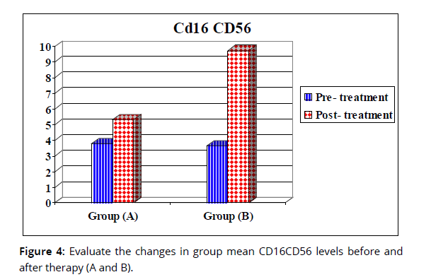
Evaluate the changes in group mean CD16CD56 levels before and after therapy (A and B)
Group (A) pre-treatment's mean SD value was 3.77± 2.37, and their posttreatment mean SD value was 5.32± 3.1; there was a statistically significant difference between these two sets of data (p= 0.0001), and the percentage change between the two sets of data was 20.56%. Group (B) pre-treatment's mean SD value was 3.65± 1.91, while their post-treatment mean SD value was 9.72 ±1.76 (Table 6). There was a statistically significant difference between these two sets of data (p= 0.0001), and the percentage of change was 83% (Figure 4).
Items |
CD16 CD56 | |||
|---|---|---|---|---|
| Group (A) | Group (B) | |||
| Pre- Treatment |
Post- Treatment |
Pre- treatment |
Post- Treatment |
|
| x̄± SD | 3.77 ± 2.37 |
5.32 ± 3.1 |
3.65 ± 1.91 |
9.72 ± 1.76 |
| MD | 1.55 | 6.07 | ||
| % of change | 20.56 % | 83 % | ||
| t-value | 4.76 | 18.66 | ||
| p-value | 0.0001 | 0.0001 | ||
| Level of Significant | S | S | ||
Evaluate the changes in group mean CD16CD56 levels after therapy (A and B)
It was shown that there was a statistically significant difference (p= 0.0001) in favor of group (B) after treatment, with mean (SD) values of 5.32± 3.1 and 9.72± 1.76 (Table 7), and the percentage of change was 82.7% (Figure 4).
Items |
CD16 CD56 (Post- treatment) |
|
|---|---|---|
| Group (A) | Group (B) | |
| x̄± SD | 5.32 ± 3.1 |
9.72 ± 1.76 |
| MD | 4.4 | |
| % of change | 82.7 % | |
| t-value | 5.52 | |
| p-value | 0.0001 | |
| Level of Significant | S | |
Discussion
Results from the current study showed that CD16 levels in children with spastic CP ranged from 58.73 to 59.85, CD56 levels from 12.8 to 13.66, and CD16 CD56 levels from 3.65 to 3.77 that indicated immunity impairment. It was explained by Sharova et al., [11] CP could be a collection of development clutters, in which the incorporate dis-regulation of innate immunity occurred as a result of non-advanced untimely brain injuries with lifelong pathophysiological. Diligent inflammation with expanded of circulating pro-inflammatory level tumor necrosis factor alpha (TNF-a) is contrarily related with recovery outcome in cerebral palsy children. On the other hand, Ortega et al., [3] added that there is immune dis-regulation coming within an innate immune response set up to fight against damaged brain cells.
Additionally, Gibson et al., [12] found that CP associated with administrative polymorphism of the β-2 adrenergic receptor causes abnormal immune responses to natural variables. Typically, confirmed by Pingel et al., [13] who used C-reactive protein (CRP) emission. It appeared 8-fold higher levels of CRP within the plasma of CP children in comparison with sound grown-ups.
Mohandas et al., [14] clarified the close relationship between immunity system and CP in infant twins whom distinguished the activation of genes related with inflammation and leukocyte-mediated immune reactions causing the improvement of CP within the influenced twin.
In the current study, the outcomes showed a significant increase in natural killer cell for expression of CD16 CD56 in children with spastic CP after applying of low level laser acupuncture on the immunity acupoints that demonstrated by Łukasiak & Jakubowski, [15] who reported that phagocytic and chemotactic parts of human leukocytes were incremented by infrared LLLT because of a case of photo biological actuation. Photo biologic cell-specific harm is additionally conceivable utilizing diode lasers on cells which have been photo sensitized and for common laser wavelength, similar to photo dynamic treatment with surface cancers. Within the immune system, LLLT has moreover appeared to function accurately and selectively.
Laser radiation at 632 and 830 nm, as reported by Lundgren et al. [16] appears to have an immune- stimulating effect.
Mustafa et al., [17] examined whether Low Level Laser irradiation influenced lymphocyte number in human entirety blood in vitro where one hundred and thirty blood samples were collected from clearly sound grown-up patients. Each experiment was split in half so that half could serve as a control and the other as a radioactive sample. Several lasers of varying wavelengths were used on the radioactive sample "405, 589, and 780 nm "with diverse fluences of 36, 54, 72, and 90 J/cm2, at a settled irradiance of 30mW/cm2. A matched understudy T- test was utilized to differentiate between non- radioactive and lighted tests. At a fluence of 72 J/cm2, the lymphocytes were measured as the greatest increment (1.6%) when compared with non-radioactive tests by employing a computerized hematology analyzer. This increment in lymphocyte number upon illumination was affirmed by stream cytometry. At a wavelength of 589 nm and fluence of 72 J/cm2, there was increasing in natural killer (NK) in expression of (CD16, CD56) cells in addition to increasing of CD45 lymphocytes, in contrast of T-suppressor (CD3, CD8) cells, T-helper (CD3, CD4) cells and CD3 T lymphocytes: there were not changes in them when differentiate with their non-radioactive partners. Out comes about clearly illustrate that natural killer cell number is modified by light, which eventually influences the entire lymphocyte check altogether.
In addition Low Level Laser with parameter of 589 nm wavelength and 72 J/cm2 fluence increments the number of human blood lymphocytes in vitro, that is called " Biological impacts of laser illumination" which are subordinate on such parameters, particularly wavelength and fluence. Outcomes illustrated that the count of NK cell is changed by illumination, which eventually influences the number of lymphocyte essentially. Laser light at 589 nm wavelength on mitochondria caused increment in this cell count that is conceivably since of the increment in intracellular ATP. The adequacy of LLLI in changing the action of cells is valuable for deciding suitable treatment for illnesses related to the immunity system.
Increasing NK cells by low-level laser acupuncture at 780nm wavelength increased immune indices in children with CP. In a similar way, Dabbous et al. [18] confirmed that low-level laser acupuncture with a wavelength of 632.8nm enhanced immunity in children with CP by raising NK cell numbers.
Yamaguchi et al., [19] during the course of the study, blood was drawn from the lower arms of seventeen healthy volunteers one hour before, and again one, two, and eight days following treatment. For five seconds, a needle was inserted into ST36 along with BL18, BL20, and BL23 as part of the treatment protocol for needling therapy. Patients' NK cell rates were found to increase dynamically and statistically significantly after eight days of needle therapy, according to the study's authors (compared to pattern). Natural killer cell counts in peripheral blood that are positive for CD16 and NCAM (CD56) indicate that these cells have multiplied. Researchers in the same study found that on days 1 and 8, the number of peripheral blood cells that produced IFN- had increased by a factor of 7 and 9, respectively. IFN- is a critical stimulant of the resistant instruments that dispose of cancer cells, and NK cells' rapid delivery of IFN- in response to "Alarm" explains why this is possible.
It was inspected by Dabbous et al., [20] who used laser acupuncture as an assistive treatment for spastic CP children. They found that laser acupuncture therapy has a useful impact on diminishing hypertonia and progressing development in these children.
However, Cherenkov et al., [21] who reported: the impact of low level laser on the action of common executioner cells from sound and tumor-bearing mice was examined. Skin within the zone of the thymus or hind limb was enlightened, the remaining body surface being screened. The light was carried out for 30 days, with the length of a single introduction being 1 min and interims between the exposures being 48 h. The impact of laser light depended on the area of the enlightened range. It was appeared that the presentation of the thymus of sound creatures for 20 and 30 days leads to a noteworthy diminish within the activity of NK cells. On the opposite, the light of the appendage for 10 or 20 days expanded the activity of natural killer cells; but when rear appendages were treated for 30 days, the action of natural killer cells diminished. While tumor development expanded the natural killer cell action, the brightening of tumor-bearing mice brought down the versatile anti-tumor resistance by diminishing the action of natural killer cells.
Novoselova et al., [22] found that the impact of introduction of low level laser at 632.8 nm wavelength and a control thickness of 0.2mW/cm2 on splenic lymphocytes and macrophages were considered the useful action. In the event that the introduction interval to did not surpass sixty seconds, the incitement in interleukin-2 (IL-2) and nitric oxide (NO) generation, in addition to increment within the action of NK cells were watched. The increment of light measurements by extension of the presentation term up to one hundred and eighty seconds initiated a noteworthy diminish in NO generation and natural killer cell activity, but IL-2 generation was not diverse from the domination level. It was watched that diminish in interferon-gamma (IFN-gamma) generation taking after laser irradiation introduction of cells for sixty or one hundred and eighty seconds, though beneath lower dosages (presentation for five or thirty seconds) IFN-gamma generation expanded. The cause of natural killer activity diminishment after 180 s looks like present a double stage effect, with initial pick up followed by postponed suppression.
Conclusion
The results of this study suggest that non-thermal low-level laser acupuncture on immune acupoint ST36 and other acupuncture points, including BL18 (Gan Shu), BL20 (Pi Shu), and BL23, can improve immunity indices in children with spastic CP without any other complaints (Shen Shu).
References
Malgorzata Sadowska,Beata Sarecka-Hujar and Ilona Kopyta (2020)."Cerebral Palsy: Current Opinions on Definition, Epidemiology, Risk Factors, Classification and Treatment Options". Neuropsychiatr Dis Treat. 16:1505-1518.
Oskoui M, Coutinho F, Dykeman J, Jetté N and Pringsheim T (2017)."Cerebral Palsy: Hope through Research".National Institute of Neurological Disorders and Stroke. July 2013.Archivedfrom the original on 21 February 2017. Retrieved21.
Ortega SB, Kong X, Venkataraman R, Savedra AM, Kernie SG, Stowe AM and Raman L (2015)."Perinatal Chronic Hypoxia Induces Cortical Inflammation, Hypomyelination, and Peripheral Myelin-Specific T cell Autoreactivity''. J. Leukoc. Biol. 99(1):21–29.
Engelhardt B and Liebner S (2014). "Novel insights into the development and maintenance of the blood–brain barrier". Cell Tissue Res. 355(3):687–699.
Sivori S, Vacca P, Del Zotto G, Munari E, Mingari MC and Moretta L.(2019)."Human NK cells: surface receptors, inhibitory checkpoints, and translational applications".Cell Mol. Immunol. 16(5):430-441.
Mustafa S, Al Musawi1 MS, Jaafar1 B, Al-Gailani M, Ahmed FMS and Nursakinah S (2016)."Effects of Low-Level Laser Irradiation on Human Blood Lymphocytes in vitro''. Laser Med. sci. 10:213.
Tony Y, Chon MJ, Mallory JY, Sara E, Bublitz A, Do P and Dorsher T (2019)."Laser Acupuncture: A Concise Review". Medical Acupuncture, 31(3).
Ezzo J, Streitberger K and Schneider A (2006)."Cochrane systematic reviews examine P6 acupuncture-point stimulation for nausea and vomiting". Journal of Alternative and Complementary Medicine, 12(5): 489–495.
Lim S (2010)."WHO standard acupuncture point locations". Evidence-Based Complementary and Alternative Medicine, 7(2):167–168.
Yim YK, Lee H and Hong KE (2007). "Electro-Acupuncture at Acupoint ST36 reduces Inflammation and regulates Immune Activity in Collagen-Induced Arthritic Mice".Evidence-Based Complementary and Alternative Medicine, 4(1):51–57.
Sharova O, Smiyan O and Borén T. (2021)."Immunological effects of Cerebral palsy and Rehabilitation Exercises in Children". Brain Behav Immun Health, 9(18):100365.
Gibson C.S., MacLennan A.H., Dekker G.A., Goldwater P.N., Sullivan T.R., Munroe D.J., Tsang S., Stewart C. and Nelson K.B.(2008)." Candidate genes and cerebral palsy: a population-based study". Pediatrics, 122(5):1079–1085.
Pingel J., Barber L., Andersen I.T., Walden F.V., Wong C., Døssing S. and Nielsen J.B.(2019)."Systemic inflammatory markers in individuals with cerebral palsy". Eur. J. Inflamm.17.
Mohandas N., Bass-Stringer S., Maksimovic J., Crompton K., Loke Y.J., Walstab J., Reid S.M., Amor D.J., Reddihough D. and Craig J.M.(2018)." Epigenome-wide analysis in newborn blood spots from monozygotic twins discordant for cerebral palsy reveals consistent regional differences in DNA methylation". Clin. Epigenet.10(1):25.
Lukasiak L and Jakubowski A (2010)."History of semiconductor". Journal of telecommunications and information technology.1:3-9.
Lundgren K, Brown M, Pineda S, Cuzick J, Salter J, Zabaglo L, Howell A, Dowsett M and Landberg G(2012)."Effects of cyclin D1 gene amplification and protein expression on time to recurrence in postmenopausal breast cancer patients treated with anastrozole or tamoxifen: a TransATAC study". Breast Cancer Research, 14 :R57.
Mustafa S, Al Musawi MS and Jaafar NS (2017). "Effects of low-level laser irradiation on human blood lymphocytes in vitro". Lasers in Medical Science, 32,405–411.
Ola A Dabbous, Morsy M, Abdelaziem FH and Said RS (2022)."Effect of laser bio-stimulation with a wavelength of 632.8 on children with spastic cerebral palsy".International Journal of Health Science,6(s4),12684-12696.
Yamaguchi N, Takahashi T, Sakura M, Sugita T, Uchikawa K and Sakaiharas S (2007). "Acupuncture regulates Leukocyte Subpopulations in Human Peripheral Blood". Advance Access Publications.4(4):447-453
Ola A Dabbous,Yousry M Mostafa,Hossam A El Noamany,Shrouk A El Shennawyand Mohammed A El Bagoury (2016)." Laser Acupuncture as an Adjunctive Therapy for Spastic Cerebral Palsy in Children"Lasers in Med.Sci .31(6): 1061–1067.
Cherenkov DA, Novoselova EG, Glushkova OV, Sinotova OA, Sultanova AN, Chudnovskii AN and Fesenko EE (2005). "The activity of natural killer cells in healthy and tumor-bearing mice after treatment with low-intensity laser light". Biofizika. Jan-Feb. 50(1):114-8. Russian. PMID: 15759510.
Novoselova EG, Cherenkov DA, Glushkova OV, Novoselova TV, Chudnovskii VM, Iusupov VI and Fesenko EE (2006). "Effect of low-intensity laser radiation (632.8 nm) on immune cells isolated from mice". Biofizika.51(3):509-18. Russian. PMID: 16808352.


