Full Length Research Article - (2023) Volume 18, Issue 4
The Effect Of Stretching Exercises By Proprioceptive Neuromuscular Facilitation Pnf Of Sensory Receptors In The Rehabili
Wafaa Sabah Mohammed Al-Khafaji* and Fatimah Hameed Kzar Al-Masoodi*Correspondence: Wafaa Sabah Mohammed Al-Khafaji, College of Physical Education and Sports Sciences, University of Baghdad, Iraq, Email:
Received: 05-Jul-2023 Accepted: 19-Jul-2023 Published: 19-Jul-2023
Abstract
The purpose of this paper is to The aim of the research is to identify the effect of stretching exercises by facilitating the neuromuscular sensory receptors (PNF) in improving the working muscle strength, range of motion, and the degree of pain for people with ankle joint injuries for team and individual sports athletes. The researchers used the experimental approach with a one-group design with two pre and post- tests. The sample was chosen by the intentional method from the players with team and individual games for young people in the National Center for the Care of Sports Talent in Baghdad Governorate, who are injured in the ankle joint and from the second degree, who are (8) players. They were distributed among the games (2 basketball players, 2 volleyball players, 2 handball players, 2 swimmers), as stretching exercises were used by facilitating neuromuscular sensory receptors (PNF) for a period of (6) weeks, with (3) rehabilitation units in the week. The researchers concluded that stretching exercises with neuromuscular facilitation of sensory receptors (PNF) have a positive effect on the rehabilitation of ankle injury for sports athletes (basketball, volleyball, handball, swimming), due to the appearance of an improvement and a clear increase in the muscle strength of the working muscles and the range of motion of the ankle joint, as well as Reducing the degree of pain in the affected area.
Keywords
Stretching exercises. Muscle strength. Ankle joint injuries
Introduction
Ankle joint injury is one of the most common injuries, because the ankle is one of the most complex joints in the body. The injury is often a rupture or stretching of the ligaments that connect the bones of the ankle, as the injury occurs as a result of various lower body movements that are not commensurate with the amount of pressure placed on this area and the size of the ankle, as well as a sudden movement, as injuries are among the main problems facing the operation. The progress of sports levels and their transfer from one level to another, it is known that injuries are always associated with physical and sports activity, and that the rate of injuries in some types of sports is higher than others, especially in sports that require players to be in contact with each other, and among these injuries is the sprained ankle In the games (basketball, volleyball, handball, swimming) that are the subject of the study, they are considered among the games that are characterized by high training load, in which the players during play, training and competitions are exposed to such an injury due to the strong friction between the players as well as the skills that the players need. In which they fall on the ankle and this exposes them to the threat of injury, as the weight of the body is heavily supported on this joint, in addition to the weakness of the ankle ligaments and the great muscular effort they are exposed to, which may prevent them from playing their games. As a result of the development of basketball and the increased interest in it as a sport to achieve good results, attention has increased in aspects of rehabilitation for injured players, as the rehabilitation aspect is very important.
Rehabilitation exercises work in the treatment and rehabilitation of sports injuries by removing the dysfunction of the affected part by taking care of weaknesses in some muscles and ligaments, developing and improving muscle strength, flexibility of the joint, the degree of neuromuscular compatibility, increasing the rate of tissue healing and the speed of getting rid of adhesions and blood calcifications that accumulate inside the joint.
Stretching by neuromuscular facilitation of sensory receptors or neuromuscular coordination (stretching using the PNF method) is one of the modern and advanced methods of training and improving flexibility, in which both stretching and contraction of the target muscle groups are involved in the work, as this method was developed initially as A form of rehabilitation and physiotherapy, and because of this function or feature, this method has high effectiveness and great effect, and one of its advantages is its targeting and focusing on specialized muscle groups, and this type of stretching exercises (PNF) is effective in increasing muscle lengthening and improving joint flexibility, As this method consists in moving the limb to the maximum possible range of motion, stabilizing the position, and then causing a steady muscle contraction of the stretched muscles, then making the muscles relax and continuing to lengthen the muscles to the maximum additional possible range of motion.
Hence the importance of the research in preparing rehabilitative exercises that carry with it the possibility of improving the strength of the working muscles, the range of motion and the degree of pain for people with ankle joint injuries for sports athletes (basketball, volleyball, handball, swimming) through the use of stretching exercises by facilitating neuromuscular sensory receptors (PNF). ), to serve our injured players in team and individual games and to assist coaches and workers in the field of rehabilitation and training.
Through the experience of the researchers being teachers of swimming, training physiology and rehabilitation of injuries, and their knowledge of the centers of sports medicine units in the treatment and rehabilitation of sports injuries, they noticed the lack of use of approaches to neuromuscular compatibility (PNF) exercises, including ankle injuries, as they relied on traditional methods of physical therapy using them devices. Therefore, the researchers decided to use stretching exercises by facilitating the neuromuscular sensory receptors (PNF) in the rehabilitation of the ankle injury of athletes.
Research Objective
Identify the effect of stretching exercises by neuromuscular facilitation of sensory receptors (PNF) in improving the strength of working muscles, range of motion, and the degree of pain for people with ankle joint injuries for team and individual sports athletes.
Research Methodology and Field Procedures
Research methodology
“The method is that intellectual organization involved in the scientific study, or it is the intellectual steps that the researcher possesses to solve a specific problem” (Malek Alshok, A. 2008). The researchers adopted the experimental approach using the one-group method, "since experimentation is a method for discovering causal relationships between phenomena" (Abedalsatar M, Hanoon W. 2009), and using the pre- and post-tests method, due to its suitability to the nature of the research problem.
Community and sample research
"The objectives that the researcher sets for his research and the procedures that he will use will determine the nature of the sample that he will choose" (Abedalsatar M, Hanoon W. (2009), as the sample was selected intentionally from the players for some group and individual games for young people in the National Center for the Care of Sports Talent in Baghdad Governorate, injured in Ankle joint and of the second degree, and the number is (8) injured players, divided as follows (2 basketball players, 2 volleyball players, 2 handball players, 2 swimmers), as the injury, its severity, and the medical history were diagnosed by the specialist doctor through a clinical examination To the injured and to determine the injury by magnetic resonance imaging (Table 1).
| No. | Variables | Measuring Unit | Mean | Std. Deviations | Median | Skewness |
|---|---|---|---|---|---|---|
| 1 | Length | Cm | 178.9 | 5.22 | 177.5 | 1.26 |
| 2 | Mass | Kg | 72.50 | 4.12 | 71 | 0.55 |
| 3 | Age | Year | 22.50 | 1.88 | 22 | 1.34 |
Methods, tools and devices used in the research
"They are the means through which the researcher can collect data and solve the problem to achieve the goals of the research, whatever those tools are like data, samples, and devices" (Siham Hassan Karim. 2009).
• Arab and foreign sources.
• Internet information network.
• Personal interviews.
• Information collection form.
• A goniometer to measure the range of motion of the ankle joint.
• The dynamometer to measure the strength of the muscles working on the ankle joint.
• A scale for measuring weight.
• Metric tape measure.
• Medical bed.
• Stopwatch.
• Camera.
• Rubber bands.
• Multiple weights.
Measurements Used in the Research
First: Measurements of muscle strength working on the ankle joint, including
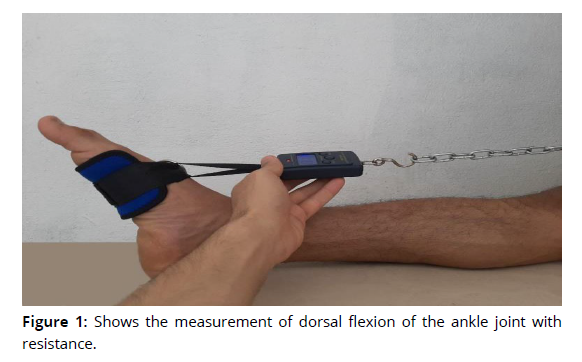
1. Dorsal flexion of the ankle joint with resistance (Reaburn andBen Dascombe . 2011).
• The purpose of the test: to measure the muscular strength of the muscles working to pull the ankle joint towards the leg.
• Tools used in the test: a Dynamometer, a metal ring to fix the device to the wall, an iron chain, a flat sitting chair, and a knee strap.
• Description of the performance: The injured person sits on a flat seated chair, as the injured player fixes the tape in the injured foot, and then the injured person performs the pulling process, after taking instructions, by fixing the heel of the foot on the flat seated chair, and the pulling process is with insteps.
• Recording: The value is calculated by reading the weight indicated by the indicator of the Dynamometer in kilograms, as it is given two attempts, and we take the best of them (Figure 1).
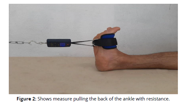
2. Pulling the back of the ankle with resistance (Reaburn andBen Dascombe .2011).
• Purpose of the test: To measure the muscular strength of the muscles working to pull the posterior ankle joint.
• Tools used in the test: a Dynamometer, a metal ring to fix the device to the wall, an iron chain, a flat sitting chair, and a knee strap.
• Description of the performance: The injured person sits on a flat sitting chair, as the researcher fixes the tape in the injured player's foot, and then the injured performs the pulling process, after taking instructions, by fixing the heel of the foot on the flat sitting chair, and the pulling process is with insteps.
• Recording: The value is calculated by reading the weight indicated by the Dynamometer in kilograms, if given to it two attempts take the best of them (Figure 2).
Second: Measurements of the range of motion of the ankle joint, including
1. Plantar flexion of the ankle joint (Ahouglum. 2012)
• The purpose of the test: measuring the range of motion of the ankle joint with the movement of the toes in the maximal direction.
• Tools used in the test: a goniometer, a sponge cushion, which the researcher used under the ankle joint, and a flat sitting chair.
• Description of the performance: the injured person sits on a flat chair, then the injured person extends the injured leg on the bench, if the leg is extended correctly, and the foot is at an angle of (90) degrees. The leg with the bones of the foot to the ankle joint, so the researcher puts the goniometer to determine the range of motion reached by the injured person during the performance of the test.
• Recording: The value is calculated by reading the angle indicated by the Goniometer, as it is given two attempts, and we take the best of them.
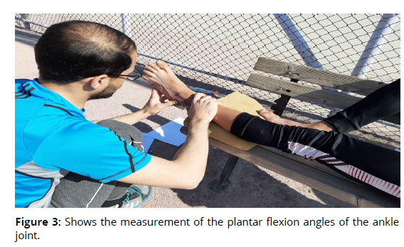
2. Dorsal flexion of the ankle joint (Ahouglum. 2012)
• The purpose of the test: to measure the range of motion of the ankle joint.
• Tools used in the test: Goniometer.
• Description of the performance: The injured person stands with his back against the wall and the injured foot is extended slightly forward from the other foot, and the injured foot is at an angle of (90) degrees so that it is standing and its fulcrum is the ankle joint. The fulcrum point, as the range of motion reached by the person with the withdrawal process is recorded by a goniometer.
• Recording: The value is calculated by reading the angle indicated by the Goniometer, as it is given two attempts, and we take the best of them (Figure 3).
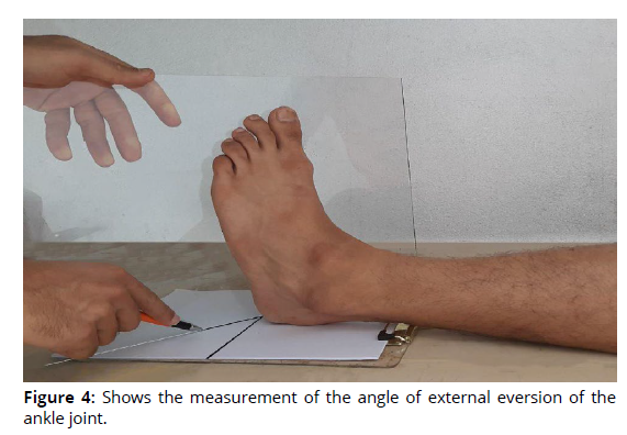
3. External eversion of the ankle joint ((Ahouglum. 2012)
• The purpose of the test: to measure the range of motion of the ankle joint by external eversion movement.
• Tools used in the test: goniometer, plate of glass (30 x 40), white paper, score pen, bench.
• Description of the performance: The patient takes a long sitting position on the ground, so when the patient hears the instruction from the therapist, he extends the injured leg towards the plate of glass, and the angle is established between the bones of the foot and the two bones of the leg and its meeting point in the ankle joint, where the patient performs the movement of the external inversion of the ankle joint By pushing a glass plate, the researcher records the angle with a goniometer, which is determined by a sheet of paper placed under the foot.
• Recording: The value is calculated by reading the angle indicated by the Goniometer, if he is given two attempts, we take the best of them (Figure 4).
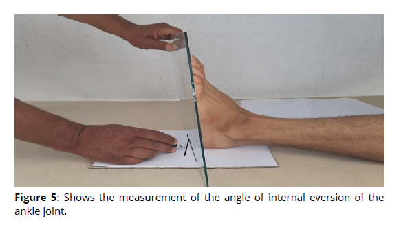
4. Internal eversion of the ankle joint (Minerva M.1990).
• The purpose of the test: to measure the range of motion of the ankle joint with the movement of internal eversion.
• Tools used in the test: goniometer, plate of glass (30 x 40), white paper, score pen, bench.
• Description of the performance: The patient takes a long sitting position on the ground, if the patient, upon hearing the instruction from the therapist, extends the injured leg towards the plate of glass, and the angles are established between the bones of the foot and the two bones of the leg and its meeting point in the ankle joint, where the patient performs the movement of the external coup To the ankle joint by pushing a glass plate, and then the researcher records the angles with a goniometer, which are determined by a paper that is placed under the foot.
• Recording: The value is calculated by reading the angle indicated by the Goniometer, as it is given two attempts, and we take the best of them (Figure 5).
Third: Measuring the degree of pain for the ankle joint (Jassim. 2015)
A form was used to measure the degree of pain from a previous study, where the motor range and resistance for each test were divided into three levels, pain is measured from the first level of (3) score if pain appears, the pain score will be recorded (3). In the first box (0) is given. For the other two squares for the same test, and if the patient does not appear to have pain at this level, a (0) is given in the square, and he moves to the second level, whose degree is (2). If pain appears, the pain is recorded (2). The third level is given for the same test (0), and if the pain does not appear in the first and second level, any sign of pain, then it is moved to the third level, which has a degree of (1). If the pain appears, the score is given, and if it does not appear, this is evidence that the motor range is normal. Then, these details are applied to the tests in the designated form, and the divisions of the motor range for each test are as follows:
First: Measurement of the plantar flexion angles of the ankle joint
• Pain when plantar flexion of the ankle joint at an angle (90 degrees) given 3 degrees.
• Pain when plantar flexion of the ankle joint at an angle (120 degrees) given 2 degrees.
• Pain when the plantar flexion of the ankle joint at an angle (135 degrees) given 1 degree.
Second: Measurement of dorsal flexion of the ankle joint
• Pain on dorsal flexion of the ankle joint at an angle of (90 degrees) giving 3 degrees.
• Pain on dorsal flexion of the ankle joint at an angle of (80 degrees) giving 2 degrees.
• Pain when dorsiflexion of the ankle joint at an angle (70 degrees) 1 degree.
Third: Measuring the angle of external inversion of the ankle joint
• Pain when the ankle joint is turned externally at an angle of (20 degrees) 3 degrees.
• Pain when the ankle joint is turned externally at an angle of (15 degrees) 2 degrees.
• Pain when the ankle joint is turned externally at an angle of (10 degrees) 1 degree.
Fourth: Measuring the angle of internal inversion of the ankle joint
• Pain when the ankle joint is internally turned at an angle (30 degrees) that gives 3 degrees.
• Pain when the ankle joint is inverted at an angle of (20 degrees) 2 degrees.
• Pain when the ankle joint is inverted at an angle of (10 degrees) 1 degree.
Fifth: dorsal flexion of the ankle joint with resistance
• Pain on dorsal flexion of the ankle joint with a weight of (10 kg) given 3 degrees.
• Pain in the dorsal flexion of the ankle joint, with a weight of (30 kg), given a degrees of 2.
• Pain on dorsal flexion of the ankle joint with a weight of (60 kg) given 1 degree.
Sixth: Pull back of the ankle with resistance
• Pain when pushing with a weight (10 kg) given 3 degrees.
• Pain when pushing with a weight (30 kg) given 2 degrees.
• Pain when pushing with a weight (60 kg) given 1 degrees.
Exploratory Experience
An exploratory experiment is considered a "mini-experiment of the main experiment, and the same conditions and conditions in which the main experiment are must be met, as far as possible, so that its results can be taken into account." (Aldulaimi, N. 2009) , the two researchers conducted the exploratory experiment on Wednesday, corresponding to 2/2/2022, on (4) of those with ankle injuries who were among the sample, before the implementation phase of the main experiment at exactly (10) am at the Medical Technical Institute in Bab Al-Moatham / Rehabilitation Branch. And with the help of the auxiliary work team, in order to see the devices and tools that will be used in the main experiment and to know the validity of their operation, in addition to conducting another exploratory experiment in the same place, to ensure the ability of the sample members to perform stretching exercises by facilitating the neuromuscular sensory receptors (PNF) in the rehabilitation of the ankle injury For athletes, in order to identify the appropriateness of the exercises for the sample and the time spent for them, and to identify the negatives and difficulties that the researchers may face during the application of the rehabilitation approach.
Pre- measurements
Pre- measurements of the research sample were taken on Friday, 4/2/2022 at exactly (10) am in the Medical Technical Institute in Bab Al-Moatham / Rehabilitation Branch, as measurements of the muscle strength working on the ankle joint, measurements of the angles of the range of motion of the ankle joint, and a measurement of the degree of the pain.
Main experience
The researchers worked on applying the rehabilitative approach in order to rehabilitate the injured in the ankle joint of the sample athletes in the games (basketball, volleyball, handball, swimming) using stretching exercises by facilitating neuromuscular sensory receptors (PNF) in rehabilitating the ankle injury of athletes, as the approach included the following:
• Rehabilitation exercises (PNF) were applied for a period of (6) weeks, at the rate of (3) units per week.
• Divide the curriculum into three levels (first, second, and third).
• The rehabilitative curriculum exercises included (18) PNF exercises, which were divided into levels for each level (6) exercises. The first level included negative exercises, while the second and third levels included exercises related to body weight and resistance exercises, as well as the use of some means such as rubber bands And the towel.
• The time of the rehabilitation unit ranged in the first and second week (10-15) minutes.
• The time of the rehabilitation unit ranged in the third and fourth week (15-20) minutes.
• The time of the rehabilitation unit ranged in the fifth and sixth week (25-30) minutes.
• The researchers followed the progression in giving stretching exercises to increase the strength and flexibility of the joint.
• Rehabilitation exercises (PNF) were implemented on Saturday 5/2/2022 and were completed on Friday 18/3/2022.
Post-measurement
Post-measurements were conducted on the research sample on Saturday 19/3/2022, and the researchers were keen to provide the same temporal and spatial conditions in the tribal measurements.
Statistical methods: The search data was processed through the Statistical Package for the Social Sciences (SPSS) (Khaleel, N. 2009).
Results and Discussion
Presentation of the results of measurements of muscular strength working on the ankle joint (Table 2).
| Measures | Measuring unit | Pre-test | Post-test | arithmetic mean of difference |
standard deviation of differences |
T value calculated | Level Sig | Type Sig | ||
|---|---|---|---|---|---|---|---|---|---|---|
| Mean | standard deviation | Mean | standard deviation | |||||||
| Dorsal flexion of the ankle joint with resistance |
Kg | 5.300 | 1.022 | 14.200 | 1.455 | 8.9 | 0.231 | 64.310 | 0.000 | Sig |
| Pull back ankle with resistance |
Kg | 7.900 | 1.791 | 18.00 | 1.516 | 10.1 | 0.324 | 33.145 | 0.000 | Sig |
Presentation the results of the range of motion measurements of the ankle joint (Table 3).
| Measures | Measuring unit | Pre-test | Post-test | arithmetic mean of difference | standard deviation of differences | T value calculated | Level Sig | Type Sig | ||
|---|---|---|---|---|---|---|---|---|---|---|
| Mean | standard deviation | Mean | standard deviation | |||||||
| Plantar flexion of the ankle joint |
Degree | 99.00 | 5.801 | 133.50 | 3.443 | 34.5 | 1.995 | 18.102 | 0.000 | Sig |
| Dorsal flexion of the ankle joint |
Degree | 82.5 | 2.331 | 70.300 | 1.455 | 12.2 | 0.883 | 16.322 | 0.000 | Sig |
| External eversion of the ankle joint |
Degree | 10.200 | 2.100 | 18.400 | 0.972 | 8.2 | 0.637 | 16.813 | 0.000 | Sig |
| Internal eversion of the ankle joint |
Degree | 17.300 | 1.987 | 28.500 | 1.733 | 11.2 | 0.299 | 42.41 | 0.000 | Sig |
Presentation the results of measuring the degree of pain (Table 4).
| Measures | Measuring unit | Pre-test | Post-test | arithmetic mean of difference | standard deviation of differences | T value calculated | Level Sig | Type Sig | ||
|---|---|---|---|---|---|---|---|---|---|---|
| Mean | standard deviation | Mean | standard deviation | |||||||
| degree of pain | Degree | 17.500 | 1.223 | 9.00 | 0.797 | 8.5 | 0.378 | 33.121 | 0.000 | Sig |
Discussion
Table 2 shows that there are significant differences between the results of the pre and post measurements and in favor of the post measurement in the muscular strength measurements represented by (dorsal flexion of the ankle joint with resistance, backward pulling of the ankle with resistance), and the researchers attribute to the effectiveness of the prepared rehabilitation curriculum and the positive effect of stretching exercises with neuromuscular facilitation of the sensory receptors (PNF), which worked to improve the muscular strength characteristic of the muscles working on the ankle joint through fixed and mobile muscle contraction exercises, as "strength development takes place by choosing fixed and moving exercises that are performed during the training curriculum to reach better results for developing the characteristic of strength" ( Jeffry e. & falkel. 1986) . The retention of the muscle in the lengthening position during the muscular contraction is related to the work of the sensory receptors within the muscle itself (muscle spindles), as "the nerve signal that forces the muscle to contract voluntarily also forces the muscle spindle to shorten with an increase in its sensitivity to the stretching reflex, which makes the muscle spindle adaptive." later" (Bead Appleton. 1998). Also, the improvement of muscular strength leads to the occurrence of neuromuscular adaptations within the muscle itself as a result of the expected training of muscle stretching exercises (PNF), which includes part of resistance exercises for a period of (6) weeks and at the rate of (3) rehabilitation units per week, as "the exercises Resistance is part of any rehabilitative curriculum to improve health and physical fitness, which depends on the number of times these exercises are in the curriculum, which ranges from one to three times. (Hameed, I., & Altay, U. 2019). As well as "the greater the number of weekly training times, the greater the rate of strength growth, as well as the intensity of the excitement of training.", the preparation of exercises is based on scientific foundations commensurate with the muscular strength of the ankle joint, as well as the nature of the muscles working on the joint and their mechanical impact, as "preparing an intensive curriculum for physical rehabilitation would help the body reach its latent fitness, while avoiding any possible complications." injury"(Minerva M. (1990).
Table (3) shows that there are significant differences between the results of the pre and post measurements and in favor of the post measurement in the measurements of the range of motion of the ankle joint represented by (plantar and dorsal flexion and external and internal inversion) of the ankle joint, and the researchers attribute the improvement in the range of motion of the various movements of the ankle joint to the effectiveness of the rehabilitation approach The stomach and the positive effect of stretching exercises by neuromuscular facilitation of sensory receptors (PNF) (neuromuscular coordination exercises) , which worked to reduce the motor limitation by increasing the range of motion of the joint in the ankle area, by increasing the special lengthening of the muscles working in the region and expanding the range of ligaments and tendons and then increasing the flexibility of the joint and thus increasing the range of motion, as the "functional effects of flexibility exercises It leads to an increase in the range of motion in the joint due to the changes that occur and the compliance of each of the muscle tendons, muscle fibers and connective tissues (Hazaa bin Mohammed Al Hazaa. 2009), and "because therapeutic exercises increase body flexibility and activity and increase neuromuscular compatibility." (Abdul Zahraa, and Farhan, A. 2022). The researchers confirm that rehabilitative exercises using (PNF) technique are very effective in increasing muscle lengthening and improving joint flexibility by adapting the sensory receptors (muscle spindles and Golgi tendons) by moving the limb to the maximum possible range of motion, stabilizing the position, and then Induce a steady muscle contraction of the tensed muscles, then make the muscles relax and continue to lengthen the muscles to the maximum possible additional range of motion (As the idea of lengthening in the (PNF) method is based on moving the limb to the maximum possible range of motion, with fixation in the position for a period of time, then causing muscle contraction to the maximum possible range of motion, and this wasp works to cause special neurological adaptation in the sensory receptors represented by the adaptation of stretch receptors In the muscle spindles to accept greater elongation, and the tension resulting from the static muscle contraction and maintaining it for a period of time leads to the activation of the Golgi tendinous bodies, which in turn activate the process of muscle contraction, which ultimately leads to an increase in muscle elongation and thus improving flexibility) ((Hazaa bin Mohammed Al Hazaa. 2009).
The researchers also confirm the use of muscle stretching (PNF) exercises for the injured in the ankle joint and applying them correctly according to the (PNF) technique led to an improvement in the lengthening of the muscles and the flexibility of the joint. By increasing the flexibility of the joints (Hussein Al-Bashtawy and Ahmed Ibrahim Al-Khawaja. 2005). Also, the use of muscle stretching exercises (PNF) in the rehabilitation curriculum depends on performing the exercises slowly and with the full range of motion of the ankle joint, "as the stretching exercises are more effective when they are performed slowly and with the full range of motion." (Al-Samarrai and Hashem Ibrahim. 1988) .Previous studies related to our current study indicate that the physiological effects of stretching exercises indicate that stretching exercises lead to an increase in the range of motion of the joint. This is due to changes in the compliance of both muscle tendons and connective tissues. 10 minutes per day for the ankle joint for a period of (6) weeks led to an increase in the range of motion of the joint by approximately (30%) and more than (50%) of the improvement occurred in the first (10) sessions ((Hazaa bin Mohammed Al Hazaa. 2009).
Table (4) shows that there are significant differences between the results of the pre and post measurements and in favor of the post measurement in measuring the degree of pain. To reduce the degree of pain suffered by the injured, as the use of muscle stretching exercises (PNF) leads to activating the injury area and then stimulating the blood circulation in the place of injury, which leads to raising the residuals of the injury and increasing the nutrition of the affected area and then its symmetry to recovery and decrease The degree of pain, as "stretching exercises (PNF) reduce the degree of pain." (Susan S. Alder, Dominiek Beckers, Math Buck. 2008), because “one of the benefits of muscle stretching is the reduction of aches and pains in the muscle.” (Arnold G. Nelson and Joko Kokonen. 2009). The optimal use of rehabilitative exercises using (PNF) technique at regular times helped the injured to significantly improve the results of the dimensional measurements that the researchers were seeking, noting that the exercises were performed slowly and carefully and mediated by specialists in the rehabilitation centers to avoid any tension that might occur in the ankle joint and thus lead to Increased pain at this stage, "as the exercises must be performed carefully to include a level that prevents the occurrence of pain, especially in the first stage of performance." (Mackenzie R. 1989) .Also, the decrease in the degree of pain is an indicator of symmetry for recovery, as with its decrease the patient can perform movements with motor ranges that are close to normal, and its presence indicates a defect, as “pain is what prevents the muscles from performing effectively and coordinating, and it can be a sign of potential damage.” (Susan S. Alder, Dominiek Beckers, Math Buck. 2008).
Conclusions and Recommendations
Conclusions
• The effectiveness of using stretching exercises by facilitating neuromuscular sensory receptors (PNF) in rehabilitating an ankle sprain injury for sports athletes (basketball, volleyball, handball, and swimming).
• The use of muscle stretching exercises (PNF) led to an improvement and a clear increase in the muscular strength of the muscles working on the ankle joint represented by (dorsal flexion of the ankle joint with resistance and pulling back of the ankle with resistance).
• The use of muscle stretching exercises (PNF) led to an improvement and a clear increase in the range of motion of the ankle joint represented by (plantar and dorsal flexion and external and internal inversion) of the ankle joint.
• The use of muscle stretching exercises (PNF) reduced the degree of pain in the ankle joint and its compliance with recovery.
Recommendations
• Adopting stretching exercises by facilitating neuromuscular sensory receptors (PNF) in the rehabilitation of ankle joint injuries for team and individual sports athletes.
• The need to put muscle-stretching exercises (PNF) within the rehabilitation curricula in the treatment centers for the rehabilitation of injuries.
• Conducting further studies of muscle stretching (PNF) exercises for upper extremity injuries represented by the shoulder joint injury.
References
Ahmed Salman Jassim. (2015). The effect of two different approaches in rehabilitating reflex ankle sprain injury for advanced handball players. Master Thesis. Karbala University. College of Physical Education and Sports Sciences.
Abdul Zahraa, S., & Farhan, A. (2022). The Effect of Musculus Rhomboideus fibromyalgia Rehabilitation Program in Youth and Advance Freestyle and Greco - Roman Wrestlers. Journal of Physical Education, 34(2), 155–171. https://doi.org/10.37359/JOPE.V34(2)2022.1264
Arnold G. Nelson and Joko Kokonen. (2009) Muscle lengthening anatomy. I 1. Kingdom of Saudi Arabia. Riyadh: Jarir Bookstore.
Siham Hassan Karim. (2009) The effect of a proposed program in developing mental visualization skills to improve the performance of the table tennis activity for the disabled. Journal of Physical Education. Baghdad University. Volume 21. Issue 1.
Malek Alshok, A. (2008). Impact exercises, especially in the development of speed and its relationship with the completion of (200 m) freestyle. Journal of Physical Education, 20(2), 259–271. https://doi.org/10.37359/JOPE.V20(2)2008.772
Fouad Al-Samarrai and Hashem Ibrahim. (1988) Sports injuries and physical therapy. i2. Jordan. Amman: Middle East Printing Company.
Qasim Hassan Hussein and Bastawisi Ahmed. Isometric muscle training. Cairo: Dar Al-Fikr Al-Arabi, 1979.
Muhannad Hussein Al-Bashtawy and Ahmed Ibrahim Al-Khawaja. (2005) Principles of sports training science. I 1. Amman: Dar Wael for publication and distribution,.
Abedalsatar M, Hanoon W. (2009). The special drills for fatness and their effect in developing some basic skills in basketball for ages (17-18). job [Internet]. Jun. 28 [cited 2023May3];21(2):363-82Availablefrom: https://jcope.uobaghdad.edu.iq/index.php/jcope/article/view/728
Aldulaimi, N. (2009). The effects of the Individual & Collective competition pattern on improving the spike and block skills and their retention in volleyball of the students. Journal of Physical Education, 21(2), 213–238. https://doi.org/10.37359/JOPE.V21(2)2009.723
Hazaa bin Mohammed Al Hazaa. (2009)Exercise physiology Theoretical foundations and laboratory procedures for physiological measurements. C 1. Saudi Arabia: King Saud University.
Bead Appleton. (1998). Stretching and Flexibility, Everything You Never Wanted to Know, by Bradford D. Appleton.
Jeffry e. & falkel. (1986). Methods of Training in sport physical Therapy, Bernhard.T. publishing editor Churchill Livingston, U.S.A., New York.
Mackenzie R. (1989).Treat Your Own Back, Orthopedic Physical Therapy Products.
Minerva M. (1990).The Journal of Sports Medicine and Physical Fitness, Jun.
Peggy Ahouglum. (2012). Examination of Musculoskeletal Injury, New York.
Peter Reaburn andBen Dascombe . (2011).Practical skills in sport and exercise science, Pearson Education Limited.
Susan S. Alder, Dominiek Beckers, Math Buck. (2008). PNF in Practice, Third Edition, Springer Mediting Verlag Heidelberg.
Khaleel, N. (2009). Loads of training to develop endurance in some physiological variables of the two league and respiratory. Journal of Physical Education, 21(1), 278–294. https://doi.org/10.37359/JOPE.V21(1)2009.713
Hameed, I., & Altay, U. (2019). The Effect of Rehabilitation Program For Hamstring Partial Tear In Advance Soccer Players. Journal of Physical Education, 31(4), 60–70. https://doi.org/10.37359/JOPE.V31(4)2019.892
